Europium »
PDB 1bub-9c8z »
2pe7 »
Europium in PDB 2pe7: Thaumatin From Thaumatococcus Danielli in Complex with Tris- Dipicolinate Europium
Protein crystallography data
The structure of Thaumatin From Thaumatococcus Danielli in Complex with Tris- Dipicolinate Europium, PDB code: 2pe7
was solved by
G.Pompidor,
J.Vicat,
R.Kahn,
with X-Ray Crystallography technique. A brief refinement statistics is given in the table below:
| Resolution Low / High (Å) | 53.68 / 1.46 |
| Space group | P 41 21 2 |
| Cell size a, b, c (Å), α, β, γ (°) | 57.550, 57.550, 149.400, 90.00, 90.00, 90.00 |
| R / Rfree (%) | 15.2 / 17.6 |
Europium Binding Sites:
The binding sites of Europium atom in the Thaumatin From Thaumatococcus Danielli in Complex with Tris- Dipicolinate Europium
(pdb code 2pe7). This binding sites where shown within
5.0 Angstroms radius around Europium atom.
In total only one binding site of Europium was determined in the Thaumatin From Thaumatococcus Danielli in Complex with Tris- Dipicolinate Europium, PDB code: 2pe7:
In total only one binding site of Europium was determined in the Thaumatin From Thaumatococcus Danielli in Complex with Tris- Dipicolinate Europium, PDB code: 2pe7:
Europium binding site 1 out of 1 in 2pe7
Go back to
Europium binding site 1 out
of 1 in the Thaumatin From Thaumatococcus Danielli in Complex with Tris- Dipicolinate Europium
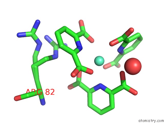
Mono view
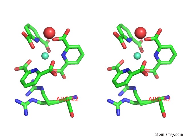
Stereo pair view
Mono view
Stereo pair view
A full contact list of Europium with other atoms in the Eu binding
site number 1 of Thaumatin From Thaumatococcus Danielli in Complex with Tris- Dipicolinate Europium within 5.0Å range:
|
Reference:
G.Pompidor,
O.Maury,
J.Vicat,
R.Kahn.
A Dipicolinate Lanthanide Complex For Solving Protein Structures Using Anomalous Diffraction. Acta Crystallogr.,Sect.D V. 66 762 2010.
ISSN: ISSN 0907-4449
PubMed: 20606256
DOI: 10.1107/S0907444910010954
Page generated: Wed Jul 31 10:40:14 2024
ISSN: ISSN 0907-4449
PubMed: 20606256
DOI: 10.1107/S0907444910010954
Last articles
Zn in 9MJ5Zn in 9HNW
Zn in 9G0L
Zn in 9FNE
Zn in 9DZN
Zn in 9E0I
Zn in 9D32
Zn in 9DAK
Zn in 8ZXC
Zn in 8ZUF